")


Description
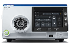
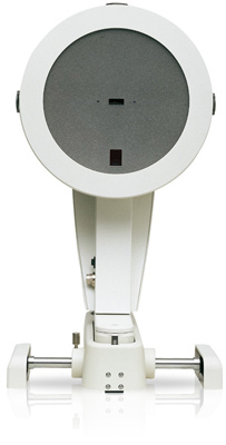
Pentacam® AXL
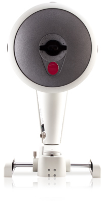
Pentacam® AXL
The new Pentacam® AXL is a systematic enhancement of the time-tested Pentacam® HR technology. In addition to anterior segment tomography, the original function of its predecessor, the Pentacam® AXL has integrated axial length measurement, a feature which allows you to make accurate IOL calculations. All using one device with one measurement procedure and reliable Pentacam® software!
MORE INFORMATION
FEATURES & BENEFITS
- High resolution cross sectional images of the anterior segment
- Optical Biometry Axial Length measurement
- IOL Calculation Display with Standard, Post Refractive, and Toric Formulas
- Axial and tangential topography maps
- Total corneal refractive power
- Pachymetry maps
- Anterior and posterior corneal elevation maps
- Zernike corneal wavefront analysis
- Anterior chamber angle measurement
- Anterior chamber depth and volume measurements
- 3D Lens Densitometry and Pentacam Nucleus Staging (automatic classification of lens opacity)
- Corneal Optical Densitometry for assessing and quantifying corneal opacification
- Fast Screening Report to quickly identifying abnormalities found in common pathologies
- Cataract Pre-op Display for premium IOLs
- Holladay Report with equivilent K readings
- Keratoconus detection and staging
- Belin/Ambrosio Enhanced Ectasia
- Contact Lens Fitting with simulated fluorescein image
- Various comparison displays including Compare 2 Scheimpflug
- Network ready – Up to 50 workstations included
- PIOL Fitting Software
- DICOM Interface
The New Pentacam® AXL offers various displays such as: Axial Length, IOL Calculation, Cataract Pre-Op, Topography maps of the anterior and posterior corneal surface, Belin/Ambrosio Enhanced Ectasia, Corneal Densitometry, Fast Screening Report, Overview of all captured Scheimpflug images, Pachymetry maps, absolute and relative, Elevation maps of the anterior and posterior corneal surface, 3D Anterior chamber analysis, Anterior segment tomography, General overview display, Keratoconus detection and classification, topometrically, Four maps, refractive, Comparative and differential analysis of two examinations, Comparison and super imposition of Scheimpflug images