")


SIZE & PHYSICAL DETAILS
X-Pod
Advanced portable imaging system
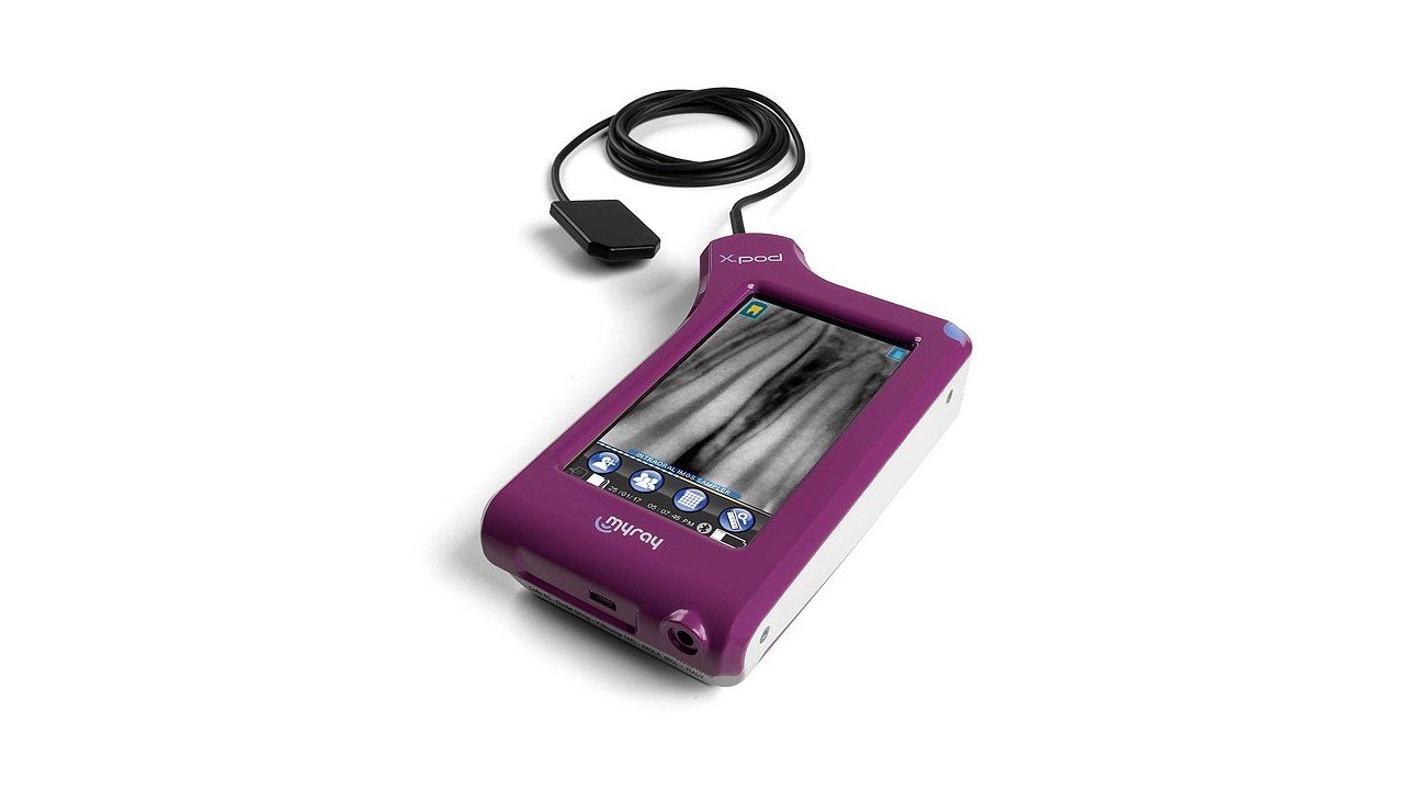
X-Pod
Perfect for your portable imaging system diagnostic needs.
Acquire, display, process and manage every detail directly in the palm of your hand on the most versatile, modern device available.
- Immediate diagnostics
- Real-time image processing
- Portability and working freedom
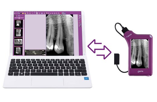
- Synchronisation with PC – iRYS software
- Bluetooth image transfer

MORE INFORMATION
FEATURES & BENEFITS
Capture, display, process and manage every detail in the palm of your hand thanks to the most versatile and modern of devices.
- HD images and hand-held processing
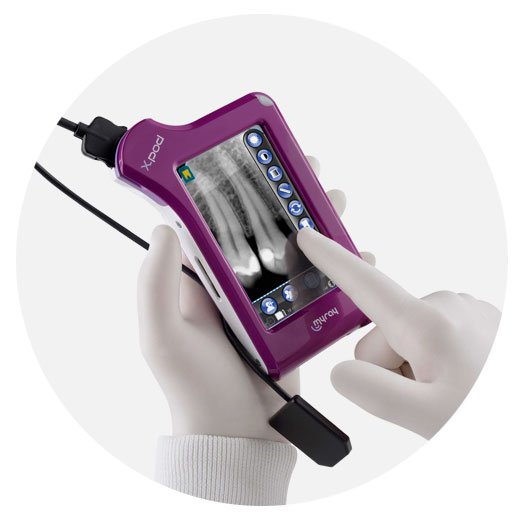
The powerful X-pod software features several advanced functions (calibration, measurement, filtering, dentition chart, rotation, patient records) with a user-friendly graphical interface to store and process images directly on the device, without any PC connection requirements.
- Self-sufficiency and portability
X-pod is compact, pocket-sized, portable, and with extra long battery power. The images are saved and organised in folders for each patient, stored on the removable Secure Digital memory card.
- Optimal workflow
Transfer and quickly synchronise data on the iRYS database of your PC via the USB cable at the end of the day, or instantly via Bluetooth.
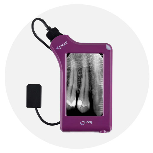
Touch-screen display
Diagnostics in the palm of your hand
Capture intraoral images, view them on the high definition, touch-sensitive display and use them for your clinical requirements. X-pod makes the workflow more efficient, improving communication with the patient and optimising your surgery’s return on investment.
The powerful X-pod software features several advanced functions with a user-friendly graphical interface to store and process images directly on the device, without any PC connection requirements.
- Edit the patient name
- Correct image rotation
- Assign a dental region on the Dentition Chart
- Measure point distances and calibrate the image
- Change filters to improve brightness and contrast
- File to patient folder
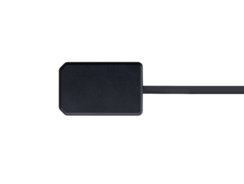
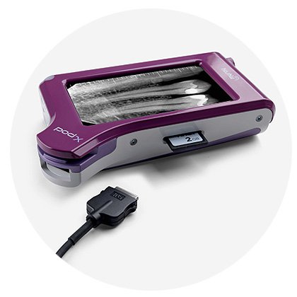
High definition sensor
Capture high definition intraoral X-ray images, view them and immediately show them to your patient for more effective communication.
The X-pod sensor consists of three different layers protected by an outer layer.
Performance
- CSL
The Caesium Iodide Scintillator intercepts the X-ray beam converting it into visible light. It is built according to a vertical growth process that generates column-shaped microstructures – capable of preserving image quality.
- FOP
This layer of optical fibres protects the sensor from direct penetration of X rays. Additionally, the vertical fibres preserve the image resolution while light propagates through the three sensor layers.
- CMOS
The CMOS capturing layer converts light into a digital image. CMOS is the latest generation of silicon digital receptors, with 20µm cells and 14bit encoding, capable of 16384 shades of grey to make sure that no detail is ever lost.
Software worklist – iRYS
Set your patient capture list from your PC with the high-performing all-in-one iRYS software and review patient records on the X-pod screen.
Capture images, view them and store them directly to patient records with correct position and dimensional information.
Transfer and quickly synchronise data on the iRYS database of your PC via the USB cable at the end of the day or instantly via Bluetooth with interference-free MyRay safe transmission technology (Patented).
Self-sufficiency and portability
X-pod is compact, pocket-sized, portable, and with extra long battery power.
The lithium-polymer battery provides enough independent power for a full day of image acquisition, at the surgery or elsewhere, without ever having to worry about charging the device. The images are saved and organised in folders for each patient, stored on the removable Secure Digital memory card.
When it is not held in the palm of your hand,
X-pod can be conveniently kept in its handy Smart Holster.
The support can be installed on any surface, such as the arm of your intraoral X-ray unit. Thanks to the efficient, adjustable control system, the image can be rotated and the display can be tilted to provide the best examination angle.