")


SIZE & PHYSICAL DETAILS
ACUSON Redwood Ultrasound System
Pushing the boundaries of imaging, performance and value
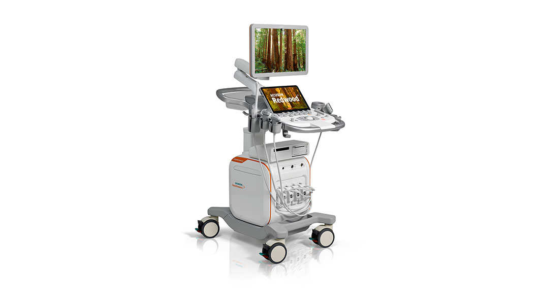
Acuson Redwood
Imaging that inspires confidence
Coherent Image Formation (CIF) for harmonic images
Using both Phase and Amplitude information to form an image, its improved alignment enables high resolution and high frame rate when compared to conventional1 ultrasound systems.
Single-crystal piezoelectric transducers for more sensitivity
Piezoelectric material is at the heart of any transducer. Single-crystal designs provide improved sensitivity and bandwidth where it’s needed most in clinical applications for both abdominal and cardiac transducers. Wider bandwidths yield better harmonic imaging, axial resolution and greater sensitivity for deeper penetration and clearer imaging.
UltraArt for greater sensitivity and quad display
Siemens Healthineers exclusive UltraArt Universal Image Processing brings you ultrasound the way you want it. Select your imaging preferences at the touch of a button using a real-time quad display. Improve the contrast resolution of different anatomical structures. Increase exam quality and consistency across different users by avoiding improper combinations of individual post processing parameters.
Reduce your Burden with Auto TEQ
AutoTEQ tissue equalization technology automatically optimizes relevant parameters so that operator adjustments are kept to a minimum. Several acquisition parameters are available, in both B-mode and pulse wave imaging modes such as gain, velocity scale, and wall filter.
MORE INFORMATION
FEATURES & BENEFITS
Advanced applications for greater clinical confidence
Meeting the demand for early detection, diagnosis and timely treatment of a variety of chronic diseases is tremendously challenging for a physician. Ultrasound imaging must enable answers to a breadth of important clinical questions — fast. To do that in the most accurate and reproducible way, the ACUSON Redwood system offers a comprehensive suite of advanced applications.
Point Shear Wave (pSWE)
Reproducible, reliable and detailed tissue stiffness information supporting liver assessment can be quickly and easily obtained using our one-touch point shear wave technology.
2D Shear Wave (SWE)
Add another dimension to quantitative shear wave imaging with color-coded shear wave maps for the 10L4 transducer in the breast and thyroid.
Strain Elastography
Virtual Touch strain elastography provides a simple and qualitative measure of lesion stiffness relative to the surrounding tissues.
Contrast-Enhanced Ultrasound (CEUS)
Contrast Pulse Sequencing (CPS) and flash sequencing technologies enable greater diagnostic confidence in the characterization of focal liver lesions.
syngo Velocity Vector Imaging (VVI)
To address the increased use of speckle-tracking echocardiography, physicians need to non-invasively assess myocardial motion and mechanics.
Stress Echo
Comprehensive and flexible stress echo package includes configurable stress echo protocols and wallmotion scoring features.
Left Ventricular Opacification (LVO)
Confidently perform LVO studies. The intuitive touch-screen layout has been streamlined to only include the functions necessary for these cardiac studies. No visual clutter—users can focus on images, not interfaces